Glaucoma, especially in the open-angle form, develops gradually, so we may simply not pay attention to the signs of the disease. Meanwhile, symptoms and treatment of increased internal eye pressure closely interconnected! The earlier the disease is diagnosed, the greater the chance that surgery can be avoided.
A special liquid constantly circulates in a person’s eyes, which has many important functions - it serves as a conductor nutrients and support for all parts of the organ. Metabolic products and bacteria are also excreted with the intraocular fluid. Normally, every minute the eye receives 2 microliters of fresh fluid and the same amount flows out through the drainage channels. If the outflow is disrupted, the pressure increases. Symptoms of increased intraocular pressure:
There are two types of glaucoma - closed-angle and open-angle. The first reason is deformation of the structure of the eye, when the iris grows and begins to compress the drainage channels. The second type is associated with deterioration of the intraocular fluid outflow system itself - weakening of vessels and channels, deposits of proteins and lipids on their walls, and weakening of muscles. Open-angle glaucoma is more dangerous, since all these processes occur very slowly; for the first few years, the disease is practically asymptomatic. On initial stage It is very difficult to detect the disease.
Symptoms that intraocular pressure is increased for open-angle and closed-angle glaucoma will be completely identical.
The opposite situations also occur when there is a deficiency of fluid in the eye. This condition has several causes, one of the main ones being general hypotension. This can also happen due to injury and water starvation. Symptoms of low intraocular pressure are as follows:
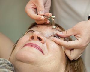
Your doctor will determine how to treat intraocular pressure disorders. Before this, it is necessary to measure exact indicators using a Maklakov tonometer.
When pressure in the eye increases, it is important to know what form of glaucoma has developed. Open angle is harder to spot, but much easier to defeat. Often when the first symptoms of increased intraocular pressure are detected, treatment is sufficient folk remedies. This is a diet aimed at reducing body weight, increasing motor activity and diuretic herbal mixtures. IN for preventive purposes It is enough to drink a course of rosehip infusion to feel better. Good results showed and special gymnastics for the eyes. With the help of exercises you can significantly strengthen  muscles and speed up metabolic processes eyes.
In most cases, surgery is prescribed, since it is not possible to influence the ocular structure in other ways. But this diagnosis is not a death sentence. In the early stages, drops may be prescribed to regulate the outflow of intraocular fluid, for example, Xalatan and Carbochol. Their action will be sufficient to prevent vision deterioration and atrophy optic nerve. Of course, you will have to use these medications on an ongoing basis for many years. But if you are categorically against eye surgery and laser correction, this option will be the only correct one.
Vision is a very important function of our body; it helps us see the world and surrounding objects. But with some problems, vision may deteriorate.
For example, its severity is affected by intraocular pressure. It can either decrease or increase. What is the normal intraocular pressure? What are the reasons for its change? And how to fix problems?
So, intraocular pressure is the pressure that is exerted on the eye wall intraocular fluid And vitreous. Normally, this indicator is constant, although minor fluctuations may be observed during the day.
But serious deviations indicate problems and can lead to undesirable consequences. So, if the value is constantly abnormal, then visual acuity will gradually decrease. A disease such as glaucoma may occur.
And if it is not treated, then vision can be lost completely and irrevocably. In addition, if the intraocular contents change its volume, this will certainly affect the shape of the eye, which means that the image will be distorted and the person will not be able to see normally.
What is the normal intraocular pressure? It is determined using special measurements and will depend on the specific method chosen. Today there are two possible:
So, the norm of intraocular pressure is now known, we can proceed to its changes. Indicators can deviate both up and down.
That is, both an increase and a decrease are possible. Both of these conditions are not normal and do not develop spontaneously. Typically, changes in the volume or composition of intraocular contents lead to certain problems, negative factors or pathology.
Increased intraocular pressure is divided into three forms:
Intraocular contents (fluid and vitreous humor) are normally contained in the eyeball in a certain volume. And this volume can change only for some reason or under the influence of certain factors.
First, let's list the reasons for increased intraocular pressure:
Now, more about the reasons for the decrease in intraocular pressure:
How will the changes manifest themselves?

Here are the symptoms of increased intraocular pressure:
Headaches may also occur (most often they are localized in the temple area).
Now in more detail about the manifestations of low intraocular pressure. They are not as obvious and noticeable as with promotion.
Often a person does not notice any changes at all and only after a year or several years does he discover that his vision has deteriorated. And yet there are some possible symptoms, rather related to related problems and pathologies that may allow one to suspect a decrease:
First of all, you need to eliminate the root cause of the change. It is important to eliminate hypertension or hypotension and get rid of other diseases ( diabetes mellitus, renal failure and so on). But also symptomatic therapy may also be required.
If there is an increase, the following measures are possible:
Here's what you can do to reduce intraocular pressure:
May your eyes be healthy!
It is common knowledge that there is a lot of fluid inside our eyes that has a certain pressure.
A condition in which the eyeball is tense and sensitive when touched closed eye, is called intraocular pressure.
Normal pressure inside the eye is approximately 15 mmHg. (within 12-20).
Intraocular pressure helps maintain the shape of the eye and the function of the eyeball. However, too high or low pressure can lead to blindness, since in the area optic disc the visual fibers are compressed. The compression blocks the flow of neurons from the retina to the optic nerves controlled by the brain.
Most dangerous disease the eye is characterized by high blood pressure (up to 60 to 70 mmHg), since this disease can lead to blindness. Intraocular pressure over 25-30 mm Hg. already poses a threat to human health and vision.
What is the nature of this type of pressure? Inside the capsule is the vitreous body, as well as the eye fluid. It is they who create a certain tension inside the capsule and tone the eye. Thanks to pressure, the normal spherical shape of the eye is maintained and normal nutrition of the eyeball is maintained. Ophthalmological disorders are always associated with disturbances in the pressure inside the eye. In such cases, vision deteriorates.
 Causes of increased intraocular pressure often include acute inflammation eyes, in which leukocytes and tissue particles block the trabecular space, resulting in increased internal tension of the eyeball.
 Reasons promotion intraocular pressure:
To do this, use a Maklakov tonometer, electrotonography or pneumotonometer. The procedure for measuring blood pressure is painless, as painkillers are used, but it is quite uncomfortable. The norm for this type of pressure is measured using a Maklakov tonometer; it is 24 mm Hg; according to a pneumotonometer, the norm is considered to be a pressure of 16-17 mm Hg.
Complications of intraocular pressure disorders include diseases such as glaucoma and visual atrophy. optic nerve. Therefore, it is imperative to start treatment in a timely manner.
In adults, the main signs include:
If drug therapy was not successful, then surgical intervention is performed, with the help of which it becomes possible to open the trabecular space and create channels to improve the flow of fluid inside the eye.
Treatment methods for this problem:
To prepare, you need to periodically, about once every 3 days, shake the container with the drink a little. You need to use this tincture every day, one and a half teaspoons, half an hour before meals.
Working at a computer for a long time can lead to illness, so you need to take breaks from work and combine mental work with physical activity. Watch out for eye strain, do eye exercises and be healthy!
We perceive 80% through our eyes. the world around us. Stressful situations, modern technologies and unfavorable environmental conditions cause irreparable damage to the eyes. The stability of the internal state of this organ changes under the influence of environmental factors, and we feel tired and uncomfortable. Internal cause These sensations may be a change in pressure inside the eye.
Fluids constantly circulate inside the eye, maintaining nutrition, hydration and normal functioning of this organ.
Fluids in the eye, in the process of natural microcirculation, can put pressure on the eye wall from the inside. This pressure is intraocular pressure (IOP). Intraocular pressure is a constant value, due to which normal size and spherical shape of the eyeball, stable vision.
In newborns, IOP is from 12-14 mm Hg. and increases by approximately 1 mmHg. 2 years before adolescence(12-13 years old).
In elderly people over 60 years of age, the IOP value should not exceed the thresholds of 10-23 mmHg. Values may vary depending on the chosen measurement technique.
The following deviations of eye pressure from the norm are observed: increased and decreased.
Increased pressure inside the eye is divided according to the duration of the disorder:
The main causes of increased pressure inside the eye:
The main reasons for decreased intraocular pressure:
At slight increase or decreased intraocular pressure, symptoms are subtle or absent. Only an ophthalmologist can detect the beginning signs of a disorder.
The main symptoms of progressive low blood pressure inside the eye:
Long-term progression of the disease can lead to a decrease in eye size.
With increased intraocular pressure, headaches, eye pain, increased eye fatigue, and temporary redness of the eyes upon strain are observed. Vision is rarely impaired.
The following symptoms are characteristic of increased IOP with a risk of glaucoma:
Diagnostics pathological changes IOP is carried out comprehensively. Consultations with a nephrologist, neurologist, neurosurgeon, cardiologist, traumatologist and endocrinologist are necessary to identify the cause of the disorder.
To diagnose changes in IOP, a number of devices and techniques are used:
After 40 years of age, intraocular pressure measurement must be performed at least once a year.
Complications that may occur with chronic change intraocular pressure level:
Treatment of all these complications is possible only surgically. In severe advanced cases, the changes are irreversible.
In case of serious deviations necessary treatment prescribed by the doctor.
The main medications for the treatment of eye hypotension are:
If pathological process found on early stage, treatment begins with direct impact on high level IOP, in this case, eye pressure drops are prescribed, which normalize metabolic processes in the tissues, promote the outflow of fluid, and reduce the pressure effect on the eye wall ( Travatan, Xalatan, Okumed, Timolol, Pilocarpine, Cosopt, Xalacom).
If drug therapy did not give results, use laser correction(iridoplasty, trabeculoplasty and others).
In advanced cases with a risk of complications, use surgical treatment(iridectomy, fistulizing interventions, operations using drains, and so on) with a hospital stay under the supervision of doctors.
For small fluctuations in IOP, to maintain visual health, they are suitable traditional methods. Oral decoctions and compresses are used as treatment.
Four lower leaves of aloe, pour 240 milliliters of boiling water, leave for 5 minutes. Strain and rub your eyes with the infusion up to 6 times per knock.
In 250 milliliters hot water Brew 15 grams of motherwort herb. Leave for an hour, strain, take 1 tablespoon 3 times a day.
Take 20 grams of dill, anise and coriander seeds and pour 500 milliliters of boiling water. Let it brew for 2 hours and strain, take 120 milliliters 3 times a day.
It stabilizes IOP well. It is necessary to drink the drink 4 times a day, 50 milliliters for 2 weeks. Then take a break for 3 weeks and repeat the course.
It will not only help remove bags under the eyes, but also quickly normalize IOP. Grate 2 raw potatoes on a fine grater, spread the mixture onto cotton pads, and place a compress on your eyes. The duration of the procedure is 15 minutes.
Collect dandelion leaves and grind in a blender. Mix a tablespoon of chopped herbs with the same volume of honey. Apply the product to the eyelids and cover with cotton swabs. Keep for 10-15 minutes.
Dilute 1 drop of peppermint oil in 100 milliliters of distilled water. Drop into your eyes.
Diet for elevated IOP provides for limiting fatty meats. The main diet in everyday nutrition should consist of products of plant origin.
With low IOP, you should adhere to the same rules, but fluid intake should be increased and foods rich in vitamin C should be added to the diet.
To maintain normal level IOP and maintaining the health of your eyes is necessary: