The materials are published for informational purposes only and are not a prescription for treatment! We recommend that you consult a hematologist at your medical institution!
The system of arteries of the head, neck and face includes large branches. They arise from the convex surfaces of the arteries that make up the aortic arch: the innominate (brachiocephalic trunk), and on the left - from the common carotid and subclavian.
The arteries of the head and neck are large vessels that arise from the aortic arch and carry blood to the organs of the neck, head and face.
At the level of the cartilage of the second rib on the right, the brachiocephalic trunk extends from the aorta after the trachea and to the brachiocephalic vein on the right. It moves to the right and upward and divides at the sternoclavicular joint on the right into 2 arteries: the right common carotid and subclavian.

Branches of the aortic arch: 1 - aortic arch; 2 - brachiocephalic trunk; 3 - left common carotid artery; 4 - left subclavian artery.
Cervical right artery shorter than the left common carotid artery by 20-25 mm. Common artery located behind the muscles: sternocleidomastoid, hyoid-scapular and muscles that cover the middle fascia of the neck. It moves vertically up to the transverse processes of the vertebrae of the neck, without dividing into branches. On top of the thyroid cartilage, both carotid arteries (right and left) are divided into internal and external with almost the same diameter.
The large subclavian artery consists of the right one, which arises from the brachiocephalic trunk, and the left one, which arises from the aortic arch. Left length subclavian artery 2-2.5 cm larger than the right one.
Important. The artery under the clavicle is responsible for the blood supply to the brain from the back of the head, the cerebellum, the brain of the back in the cervical region, the muscles and organs of the neck (partially), the shoulder girdle and the upper limb.

Photo 2 shows the dislocation of the arteries of the head and neck:

The arteries of the head, neck and face transport blood, nutrients: trace elements, vitamins and oxygen to controlled areas. Let's take a closer look.
The paired artery extends into the sternocleidomastoid muscle, omohyoid muscle, trachea, esophagus, pharynx and larynx. The endings of the artery are located in the carotid triangle, next to the thyroid cartilage of the larynx, where the branches are divided into external and internal - the terminal carotid arteries.
It stretches along the carotid and submandibular triangle, the submandibular fossa (inside the parotid gland). Consists of anterior, posterior, medial and terminal groups of branches. Ends with two terminal branches near the neck lower jaw.

Anastomosis with the mental artery.
Anastomosis occurs between: ascending palatine and descending palatine, ascending pharyngeal arteries; submental and sublingual; angular and dorsal nasal (from the ophthalmic) artery.

The function of blood supply to the soft tissues of the face is performed by the branches of the arteries:
The orbit is supplied with blood by arteries: the ophthalmic (branch) and the middle meningeal (branch of the maxillary artery) through the lacrimal artery of the anastomatic branch.

The oral cavity is supplied by the lingual branch, which belongs to the external carotid artery. The hypoglossal branch refers to the lingual artery, which belongs to the external carotid. The cheeks and lips are supplied with blood by the facial artery. Bottom oral cavity and the area under the chin is fed by the submental artery (from the facial branch). The floor of the oral cavity is supplied with blood from the mylohyoid branch (from the inferior alveolar artery). The mucous membrane of the gums is supplied with blood by the alveolar artery with dental branches. The cheeks are supplied with blood by the buccal branch of the maxillary artery.
Blood flows to the maxillary gums from the anterior superior alveolar arteries. Blood flows to the palate, tonsils and gums from the descending palatine artery, a branch of the maxillary artery. The blood supply to the tongue is carried out by arteries: the lingual (external carotid branch) and the facial (tonsilal branch).
The salivary glands are supplied with blood by arteries:
The nasal cavity is supplied by arteries: anterior ethmoidal, posterior ethmoidal (branches ophthalmic artery), posterior lateral nasal (branches of the palatine sphenoid artery), posterior artery of the nasal septum (branches of the palatine sphenoid artery).
The maxillary teeth receive blood from the arteries: the posterior and anterior superior alveolar. The mandibular teeth are supplied with blood from the inferior alveolar artery.
Among diseases of the arteries of the head, neck, and face, the following are considered dangerous:
They are characterized by protrusion of the walls of the arteries and the absence of their three-layer structure. When a cerebral aneurysm ruptures, subarachnoid hemorrhage is possible with blood penetrating into the subarachnoid space of the brain.
An aneurysm can be arteriovenous or arterial and often occurs at the site of arterial branching. The shape is: saccular aneurysm (for example, anterior communicating artery, bifurcation of the middle cerebral artery), internal fusiform and fusiform.
Narrowing of the cervical arteries and brain or atherosclerosis is accompanied by frequent attacks unbearable headache, which reduces memory. The vessels narrow when they are deposited and accumulated on the walls. cholesterol plaques, reducing the clearance. The speed of blood flow decreases, so the vessels pass less blood, and with it nutrition and oxygen.

Important. Atherosclerotic plaques are formed in cracks in the walls of arteries during their pathological conditions. They lose their elasticity with an increase in cholesterol levels in the blood, which leads to the appearance of cracks.
Plaques attract platelets, which promote blood clotting and the formation of blood clots. With acute narrowing of blood vessels, a stroke can occur, speech is impaired and vision decreases. A pre-infarction condition, cerebral infarction or hemorrhage is possible if blood circulation is suddenly disrupted.
Hypoplasia (often congenital) of the vertebral artery impairs hemodynamics (blood circulation), especially in the posterior parts of the brain. This leads to dysfunction of the heart and circulatory system, internal organs And vestibular apparatus. To diagnose and check the artery, study it functional state, roundabout blood flow, angiography is performed - contrast X-ray examination. At the same time, they will find out how long the pathological process has lasted.
When blood flow in two, right or left, vertebral arteries is weakened, blood circulation in the central nervous system worsens. These arteries provide 30-32% of the blood flow to the brain. With osteochondrosis, blood flow decreases and posterior cervical sympathetic syndrome occurs, with symptoms similar to migraines. For diagnosis, carry out Doppler ultrasound, neck X-ray, MRI.
If cervical artery syndrome is confirmed, treatment is aimed at eliminating dizziness, darkening of the eyes, headache, hearing and visual impairment and arterial hypertension.
Important. Measure the velocity of the middle cerebral artery for comparative assessment indicators of fetal blood flow speed, if pregnant women have Rh immunization, gave birth to children with Rh (-) and Rh (+) blood, in the fetus or newborn varying degrees hemolytic disease.
Using ultrasound and Doppler blood flow in the middle cerebral artery of the fetus, you can easily diagnose the severity of HDP in case of Rhesus conflict, fetal diseases affecting hemodynamics, including anemic syndrome, and study the fetal blood circulation in dynamics, without using invasive technologies.
The role of the brain in human body difficult to overestimate. Our lives are subordinated to Him, not only in psychologically, but also regarding physiology. Optimal functioning of every organ in the body, every muscle, continuous processing of information coming from the outside world, correlation of images of the desired result and the existing reality, regulation of behavior on the way to achieving goals - about the functions human brain As the crown of evolution, a lot can be said.
 In order for this entire mechanism to carry out its work without failures, it is necessary large number energy provided by oxygen and nutrients. Brain tissue requires approximately twenty times more resources than muscle tissue of similar mass. And in case oxygen starvation the brain will suffer first of all due to its special sensitivity.
Review from our reader Victoria Mirnova
I’m not used to trusting any information, but I decided to check and ordered a package. I noticed changes within a week: constant pain in my heart, heaviness, pressure surges that tormented me before receded, and after 2 weeks disappeared completely. Try it too, and if anyone is interested, below is the link to the article.
For this reason, the brain is equipped with an excellent system blood vessels. After all, it is the blood that brings to all the necessary organic cells of the body, minerals, as well as oxygen and hormones.
The vessels of the brain are formed by the paired internal carotid artery and the unpaired basilar artery. Quite large and branched, these main arteries cover the brain, including the cerebellar region, as well as top part spinal cord. They also bring blood to the neck and other organs of the head.
But nature has thought out a backup plan in case of blood clots or other vascular pathologies, capable of placing blood circulation through these vessels. All cerebral arteries unite approximately in the area of the brain stem, forming the Circle of Willis. And from this circle, in turn, emerge the three main paired arteries of the brain.
If we consider the surface of the cerebral hemispheres, we can distinguish three arteries:

The arteries in the brain and neck are a network of branched vessels. Many thin arterial trunks penetrate all layers of the medulla.
But if such a functional influx is formed for useful substances, a system for removing already used resources, waste products, and carbon dioxide from the brain is also necessary. The veins of the head serve this purpose, located, like the arteries, in the upper and lower layers of the brain.
 But the so-called venous collectors pass, among other things, between the layers of the dura mater. They are called sines. Their distinctive feature against the background of all other veins is the absence of valves and muscularis propria. They are rigid in structure. This is necessary for better outflow venous blood. This also causes strong venous bleeding if a person receives a head injury.
All arteries and veins of the head and neck are interconnected by anastomoses, that is, intervascular connections. These connections play a fairly important role physiologically, since they serve as a tool for adapting blood circulation in case of possible pathologies.
Anastomoses are of the following types:

The arteries and veins of the head and neck must be adapted to possible changes in environmental conditions inside the body, therefore their structure has a three-level structure. Each layer has its own purpose.
Artery wall structure:

The structure of the vein wall also consists of three levels, but there are some differences. This is mainly due to the fact that there are two types of veins: muscular and non-muscular. The latter are localized in the retina, hard meninges, placenta, splenic trabecula, bone substance. Often the outer layer is fused with the tissue of the organ in which the amuscular vein is located, which prevents collapse.
To clean VESSELS, prevent blood clots and get rid of CHOLESTEROL - our readers use the new natural preparation, which is recommended by Elena Malysheva. The product contains blueberry juice, clover flowers, native garlic concentrate, rock oil, and wild garlic juice.
In muscle veins, smooth muscle cells are present in all three layers of blood vessels. Muscular elements can be developed to varying degrees: weak, medium and strong. This is largely due to the location, as well as the size of the vein.
In inner layer The vein structure also contains muscle cells. But there are no elastic membranes, and along the entire length of the vessel semilunar valves are located at a sufficient distance from each other. Their purpose is to prevent blood from flowing back.
Many of our readers actively use the well-known method based on Amaranth seeds and juice, discovered by Elena Malysheva, to CLEAN VESSELS and reduce CHOLESTEROL levels in the body. We recommend that you familiarize yourself with this technique.
Do you still think that it is completely impossible to RESTORE blood vessels and the BODY!?
Have you ever tried to restore the functioning of your heart, brain or other organs after suffering pathologies and injuries? Judging by the fact that you are reading this article, you know firsthand what it is:
Did you know that all these symptoms indicate INCREASED CHOLESTEROL levels in your body? And all that is necessary is to bring cholesterol back to normal. Now answer the question: are you satisfied with this? Can ALL THESE SYMPTOMS be tolerated? How much time have you already wasted on ineffective treatment? After all, sooner or later the SITUATION WILL GET WORSE.
That's right - it's time to start putting an end to this problem! Do you agree? That is why we decided to publish an exclusive interview with the head of the Institute of Cardiology of the Ministry of Health of Russia - Renat Suleymanovich Akchurin, in which he revealed the secret of TREATING high cholesterol.
Our brain functions normally due to circulatory system, which is involved throughout the body. The anatomy of the vessels of the head and neck includes the main arteries that supply both organs, many small vessels and capillaries. But the most important are the carotid arteries, which have their own branches. This is an external vessel that nourishes cervical region both the front part and the internal part, feeding the cranial cavity and eye sockets.
To understand how blood circulates, you need to have at least the slightest knowledge about anatomical structure vascular systems. So, the blood moves in the upper cycle through the brachiocephalic arteries, carotid and left subclavian, after which it is cleared directly into the jugular and subclavian veins.
The largest and first is the brachiocephalic artery, which forms the right carotid and right subclavian arteries. Thanks to major artery Almost the entire body, and in particular the brain, is supplied with blood through the vertebral vessels. And they, in turn, supply oxygen to the entire circulatory system of the neck and brain.
The carotid arteries are conventionally divided into internal and external vessels. Vessels of the head: anatomy of the external and internal carotid arteries:
Blood Vessels of the Head and Neck: Anatomy involves the flow of blood within the skull through the jugular vein, which flows from the right and left sides from the scalp, parotid gland and facial muscular system into the subclavian vein.
The subclavian vessels supply blood to almost the entire body and the brain system of the head and back. They have left and right arteries, and the right one is 4 cm shorter than the left one. There is a transverse vessel of the neck in the subclavian artery, while the right and left branch intertwined with the external carotid artery, axillary and subscapular vessels. This provides a unique opportunity to redirect blood flow to another direction, which is called collateral circulation.
The anatomy of the vessels of the head and neck includes the BBB, that is, the blood-brain barrier, consisting of a semi-permeable membrane. It is this membrane that controls the entire capillary system. As is known, the capillaries of the circulatory system are lined with endothelial cell formations, but in different parts of the body in different ways. For example, throughout the body, except for the brain, cells have small spaces between each other. And in the brain region, the cells on the capillaries are in close contact with each other, thereby creating the tightest possible connection. Endothelial cells are connected with the help of glial cells (astrocytes), due to which a reliable protective barrier is created around all vessels. In addition, astrocytes help transport electrolytes directly from the brain into the blood fluid.

Particular attention should be paid to the common carotid artery, which is a steam artery. For example, the right artery originates at the bifurcation site in the innominate artery behind the clavicular joints. At the same time, it is tied mainly to the neck. But the artery on the left side begins behind the innominate vessel, but in the upper section of the aortic arch. Thus, the left carotid artery belongs to the thoracic and cervical parts.
Both carotid arteries in the cervical region emerge from under the sternum and clavicular joint, rising obliquely upward. And already on upper limit The thyroid cartilage is divided into external and internal carotid artery. As a rule, these arteries do not give off branches, but in some cases the vessel can give off branches to the vertebral veins, larynx, thyroid gland or pharynx.
The ASG, left CCA and RCA emerge from the aortic arch. At the level of the right sternoclavicular joint, the ASG is divided into the right CCA and RCA.
The RCA lies in an arc on the dome of the pleura, passes between the anterior and middle scalene muscles, and dives from under the collarbone into the armpit.
Click on pictures to enlarge.
The OCA passes behind the sternocleidomastoid muscle. The CCA has no branches; at the upper edge of the thyroid cartilage it is divided into the ECA and the ICA.
The extension of the bifurcation (bulb) contains chemo- and baroreceptors that regulate the functioning of breathing, heart and blood vessels.

The ECA begins medially, then runs outward from the ICA; has a short trunk; near the angle of the lower jaw it divides into eight branches.
Branches of the ECA: superior thyroid, lingual, facial, ascending pharyngeal, occipital, posterior auricular, maxillary, superficial temporal.
The BSA is wider than the NSA; on the neck it rises between the pharynx and the IJV, does not give branches; passes into the cranial cavity through the canal of the pyramid of the temporal bone.
In the skull, the branches of the ICA are the ophthalmic, anterior cerebral, middle cerebral, posterior connective; maxillary artery - middle meningeal.

The VA departs from the first segment of the RCA, ascends through the openings of the transverse processes C6-C1, and enters the skull through the foramen magnum.
The PAs of both sides merge into the main artery at the posterior edge of the bridge; the basilar artery divides into the posterior cerebral artery leading edge bridge.
I segment from the mouth to C6; II segment in the canal of the transverse processes C6-C2; III segment from C2 to the entrance to the skull; IV segment before merging into the basilar artery.

The ICA and VA form an arterial circle at the base of the brain with the help of the anterior and posterior communicating arteries; more often one of the branches is missing.

Assessment of cerebral blood flow includes the brachiocephalic arteries at the level of the neck and the intracranial vessels of the brain.
They use a 3-5 MHz convex or sector sensor, as well as a 7-18 MHz linear sensor.
Lying on your back, neck extended, head slightly turned in the opposite direction. There are 5 minutes of rest before the test.
Three approaches to the arteries of the neck: anterior - in front of the sternocleidomastoid muscle, lateral - along the CM, posterior - behind the CM.
In B-mode and CDC, the CCA is scanned up to the bifurcation; above the bifurcation, the ECA is examined through an anterior approach, and the ICA through a lateral approach.
Using a 3-5 MHz convex or sector sensor, the course of the arteries emanating from the aortic arch is examined - PGS, RCA, CCA, ECA and ICA to the entrance to the skull, as well as VA from the mouth to the entrance to the skull.
The course of the vessels is normally rectilinear, but there is tortuosity - C-, S- bends, loops. Up to 12 years of age, disruption of the course can be considered as a reserve of vessel length necessary during the period of intensive growth.
A 7-18 MHz linear sensor is used to examine arterial walls and the Doppler spectrum.
The vessel wall is examined with a 7-18 MHz linear sensor. When the ultrasound beam is directed at 90°, the reflection and contrast of the image are maximum.
IMTs comprise the intima and media of the vascular wall. The adventitia merges with the surrounding tissues. IMT of the CCA and ICA is measured 1 cm below and above the bifurcation.
The intima is represented by endothelium and subendothelium; media - in the CCA there is predominantly elastic stroma, in the ICA with a pronounced muscle component.
IMT is better visible on the distant wall - an anechoic media between the hyperechoic intima and adventitia. Normally 0.5-0.8 mm, in older people 1.0-1.1 mm.

In M-mode, the diameter of the vessel is measured between the intima and adventitia in systole and diastole.

Evaluate distal section PGS, RCA, CCA throughout, ICA from the mouth to the entrance to the skull, ECA in the initial segment, VA in segments V1 and V2.
To study the PGS, the sensor is placed in the jugular notch, and the beam is directed to the right. The ASG is divided into the right RCA and CCA. The ostium of the left CCA and RCA is too deep to see.
RCA segment I is examined above the sternoclavicular joint, segment II - above the clavicle, the beam is directed downwards, segment III - under the clavicle.
To study the CCA, the sensor is placed along the outer or inner edge of the sternocleidomastoid muscle. The CCA is assessed along its entire length from the mouth to the bifurcation.
At the base of the neck medially to the CCA thyroid gland, outside - inside jugular vein. Under the pressure of the sensor, the IJV is compressed, but the CCA is not.

From the base of the neck, move the sensor upward to the bifurcation of the CCA - the site of division into the ECA and ICA. There is a small extension here - an onion.

At the bifurcation of the CCA, the bulb expands, the bare trunk of the ICA and the branching ECA begin. The first branch of the ECA is the superior thyroid artery.

At the bifurcation level, the ICA is wider than the ECA; located outward and posterior to the ECA, above it moves inward; has no branches on the neck.

In the bulb, the laminar flow along the main axis of the ICA is red, and the turbulent flow zone at the outer wall is colored blue.
Located outside the bulb nerve plexus and carotid body. In rare cases, a tumor of the carotid body occurs.

The difference between the ECA and the ICA: at the bifurcation level, in 95% of cases the ECA is located medially; the diameter of the ESA is smaller; Small branches extend from the ECA on the neck.

The PA is scanned longitudinally inwards from the sternocleidomastoid muscle, from the angle of the lower jaw to the upper edge of the clavicle.
PA is characterized by asymmetry, usually the left is larger than the right. When PA is less than 2 mm, we can talk about hypoplasia.
To study the first segment of the VA, the sensor is moved to the clavicle. Normally, the VA leaves the RCA at the level of C7 and enters the bony canal at the level of C6.
Options are possible: the left VA arises from the aortic arch and enters the bony canal at the C5 level.
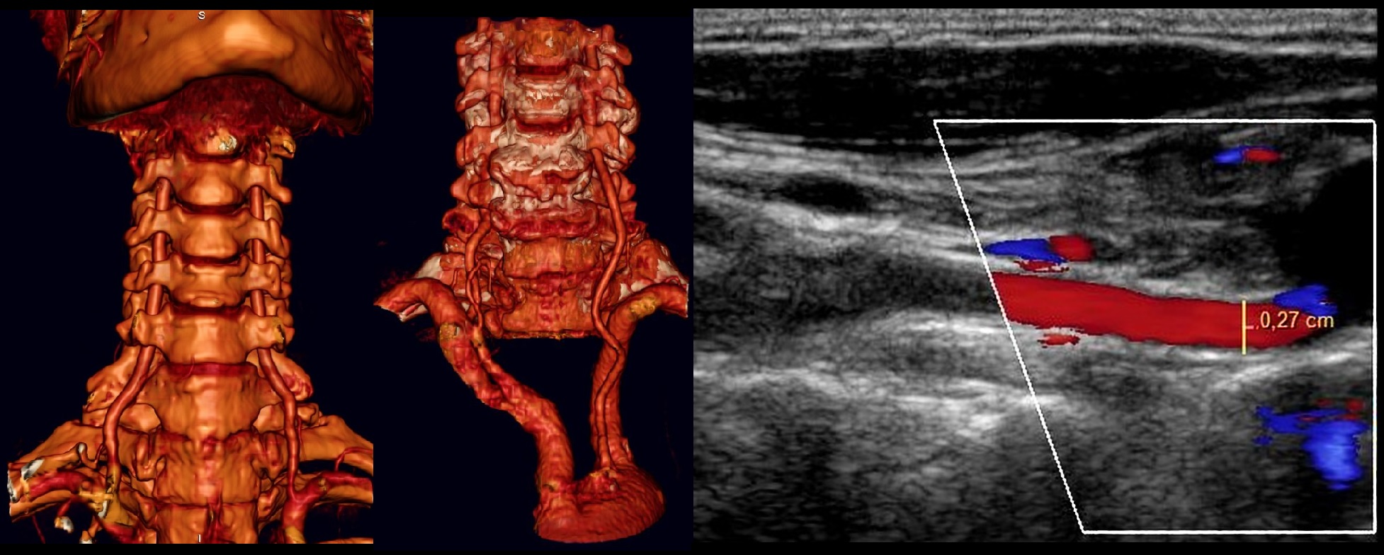
II segment of the PA has an intermittent appearance, because passes in the bone canal of the transverse processes C6-C2 and acoustic shadowing occurs at the site of the transverse processes.
If the blood flow speed in adjacent areas is approximately the same, it means pathological changes not in the blind spot.
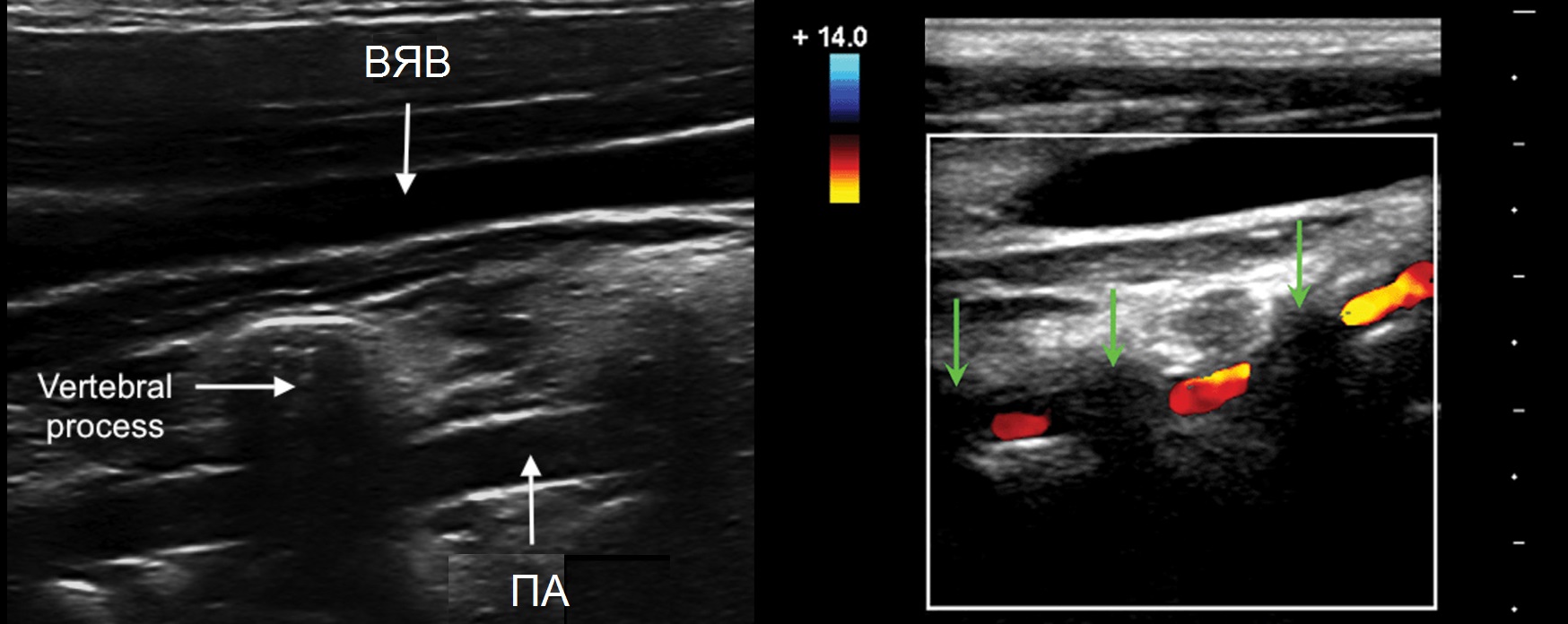
For PA segment III, a convex sensor may be useful; Due to physiological deformation, it is impossible to correctly assess blood flow.
For segment IV, the VA is examined with a 1.5-2.5 MHz sector sensor through the foramen magnum with the patient in the prone position.

Read the basics of triplex scanning. Normal indicators in the vessels of the neck and head in adults and children, see.
The CCA spectrum has a sharp rise and a narrow peak in systole, a low flow in diastole, and a dicrotic notch in late systole and early diastole.

The spectrum of ESA is similar to OSA, sometimes there is a retrograde flow in diastole, a “shooting” sound. Tap on the surface temporal artery, you will see T-waves on the NSA spectrum.

The ICA spectrum has a gradual rise and a wide peak in systole, a high antegrade flow in diastole, almost no pulsation, and a “blowing” sound.

The spectrum of segment II of the VA is similar in shape to the ICA, Vps and Ved are 1.5 times lower, the flow is exclusively antegrade. Vps may decrease in the upper sections, but not more than 20%.
The VAs of both sides have the same lumen only in 26-44% of cases, velocity asymmetry is often determined, in healthy<20%.

The ICA and VA carefully nourish the brain, the physiological deformation in the III segment of the arteries smoothes out the pulsation.
In the CCA and ECA there is high resistance, low flow in diastole; in the ICA and VA there is low resistance, high flow in diastole.

Blood flow velocities and indices are compared with normal values. On the vessels of both sides, the asymmetry of Vps should not exceed 20%, RI and PI indices - 10%.
Take care of yourself Your Diagnosticer!
The neck is a part of the human body that connects the body and head. Despite its small size, it contains many important structures, without which the brain would not receive the blood necessary for functioning. Such structures are the vessels of the neck, which perform an important function - the movement of blood from the heart to the tissues and organs of the neck and head, and then vice versa.
In the front of the neck there are paired carotid arteries and the same paired jugular veins.
It is divided into right and left, located on opposite sides of the larynx. The first one arises from the brachiocephalic trunk, so it is slightly shorter than the second one, which arises from the aortic arch. These two carotid arteries are called the common carotid arteries, and they account for 70% of the total blood flow going directly to the brain.
The internal jugular vein runs next to the CCA, and the vagus nerve is located between them. The entire system consisting of these three structures makes up the neurovascular bundle of the neck. Behind the arteries is the cervical section of the sympathetic trunk.
OCA does not produce branches. And upon reaching the carotid triangle, approximately at the level of the 4th cervical vertebra, the internal and external are divided. On both sides of the neck. The area where bifurcation occurs is called bifurcation. This is where the artery dilates - the carotid sinus.
On the inside of the carotid sinus is the carotid glomus, a small glomerulus rich in chemoreceptors. It reacts to any changes in the gas composition of the blood - the concentration of oxygen, carbon dioxide.
Located closer to the front surface of the neck. During its movement up the neck, the NSA gives off several groups of branches:
The terminal branches of the ECA are divided into even smaller vessels and supply blood to the thyroid, salivary glands, occipital, parotid, maxillary, temporal regions, as well as facial and lingual muscles.
Performs the most important function in the general blood flow, which is provided by the vessels of the head and neck - blood supply to a larger area of the brain and the human visual organ. It enters the cranial cavity through the carotid canal and does not produce branches along the way.
Once in the cranial cavity, the ICA bends (damper), penetrates the cavernous sinus and becomes part of the arterial circle of the cerebrum (circle of Willis).
Branches of the ACA:
These neck vessels carry out the reverse process - the outflow of venous blood. There are external, internal and anterior jugular veins. Blood enters the external vessel from the back of the head closer to the ear area. And also from the skin above the shoulder blade and from the front area of the face. Going lower, not reaching the clavicle, the IAV connects with the internal and subclavian. And then the inner one develops into the main one at the base of the neck and bifurcates into right and left.
The largest main vessel of the cervical spine is the IJV. It forms in the area of the skull. The main function is the outflow of blood from the vessels of the brain.
Most branches of the jugular veins bear the same names as the arteries. With those arteries that accompany it - lingual, facial, temporal... the exception is the mandibular vein.
In the area of the cervical spine there is another pair of arteries - the vertebral ones. They have a more complex structure than the carotid ones. They depart from the subclavian artery, follow behind the carotid arteries, and penetrate in the area of the 6th cervical vertebra into the canal formed by the openings of the transverse processes of the 6th vertebrae. After leaving the canal, the vertebral artery bends, passes along the upper surface of the atlas and enters the cranial cavity through the large posterior foramen. Here the right and left vertebral arteries merge and form a single basilar artery.
The vertebral arteries give off the following branches:
The basilar artery also forms a group of branches:
The anatomy of the vertebral arteries allows them to supply the brain with 30% of the blood it needs. They supply the brain stem, occipital lobes of the hemispheres and the cerebellum. This entire complex system is usually called vertebrobasilar. “Veterbro” – associated with the spine, “basilar” – with the brain.
The vertebral vein begins at the occipital bone - another of the vessels of the head and neck. It accompanies the vertebral artery, forming a plexus around it. At the end of its path in the neck it flows into the brachiocephalic vein.
The vertebral vein intersects with other veins of the cervical spine:
The anatomy of the vessels of the neck and head also includes lymphatic vessels that collect lymph. There are deep and superficial lymphatic vessels. The first ones run along the jugular vein and are located on both sides of it. The deep ones are located in close proximity to the organs from which lymph flows.
The following lateral lymphatic vessels are distinguished:
Deep lymphatic vessels collect lymph from the mouth, middle ear, and pharynx.
The nerves of the neck also perform an important function. These are diaphragmatic, muscular and skin structures located at the same level as the first four vertebrae of the neck. They form nerve plexuses from the cervical spinal nerves.
Muscle nerves are located close to the muscles and supply impulses for neck movements. Diaphragmatic ones are needed for movements of the diaphragm, pleura and pericardial fibers. And the cutaneous ones produce many branches that perform individual functions - the auricular, occipital, supraclavicular and transverse nerves.
The nerves and vessels of the head and neck are interconnected. Thus, the carotid artery, jugular vein and vagus nerve form an important neurovascular bundle of the neck.

The vessels located in the neck area are susceptible to many pathologies. And they often lead to a disastrous result – ischemic stroke. From a medical point of view, a narrowing of the lumen in blood vessels caused for any reason is called stenosis.
If pathology is not detected in time, a person may become disabled. Because the arteries in this area supply blood to the brain and all tissues and organs of the face and head.
Although there are many reasons for the pathological narrowing of the lumen, the result is always the same - the brain experiences oxygen starvation.
Therefore, with vascular disease in the neck, the symptoms look the same:
Diseases that provoke narrowing of the lumen in the cervical vessels:
As a rule, the vertebral arteries are exposed to external influence. Because they are located in a vulnerable area. Abnormal development of the vertebrae, muscle spasm, extra rib... Many factors can affect the vertebral arteries. In addition, incorrect posture during sleep can cause compression.
Tortuosity is also characteristic of the vertebral arteries. The essence of this disease is that elastic fibers predominate in the tissues that make up the vessels. And not the required collagen ones. As a result, their walls quickly become thinner and curl. Tortuosity is hereditary and may not manifest itself for a long time. Atherosclerosis can provoke tortuosity.
Any anatomical defect of the arteries is dangerous not only for human health, but also for his life. Therefore, if the slightest symptoms appear, you should consult a doctor. Don't wait for the disease to progress.
To make a correct diagnosis, doctors resort to various examinations.
Here are some of them:
The method of treating vascular diseases is selected individually for each patient.
And, as a rule, it consists of the following activities:
Treatment should be comprehensive and take place under the strict supervision of a doctor.
The anatomy of the neck has a complex structure. Nerve plexuses, arteries, veins, lymphatic vessels - the combination of all these structures ensures the relationship between the brain and the periphery. A whole network of vessels supplies all tissues and organs of the head and neck with arterial blood. Be attentive to your health!


